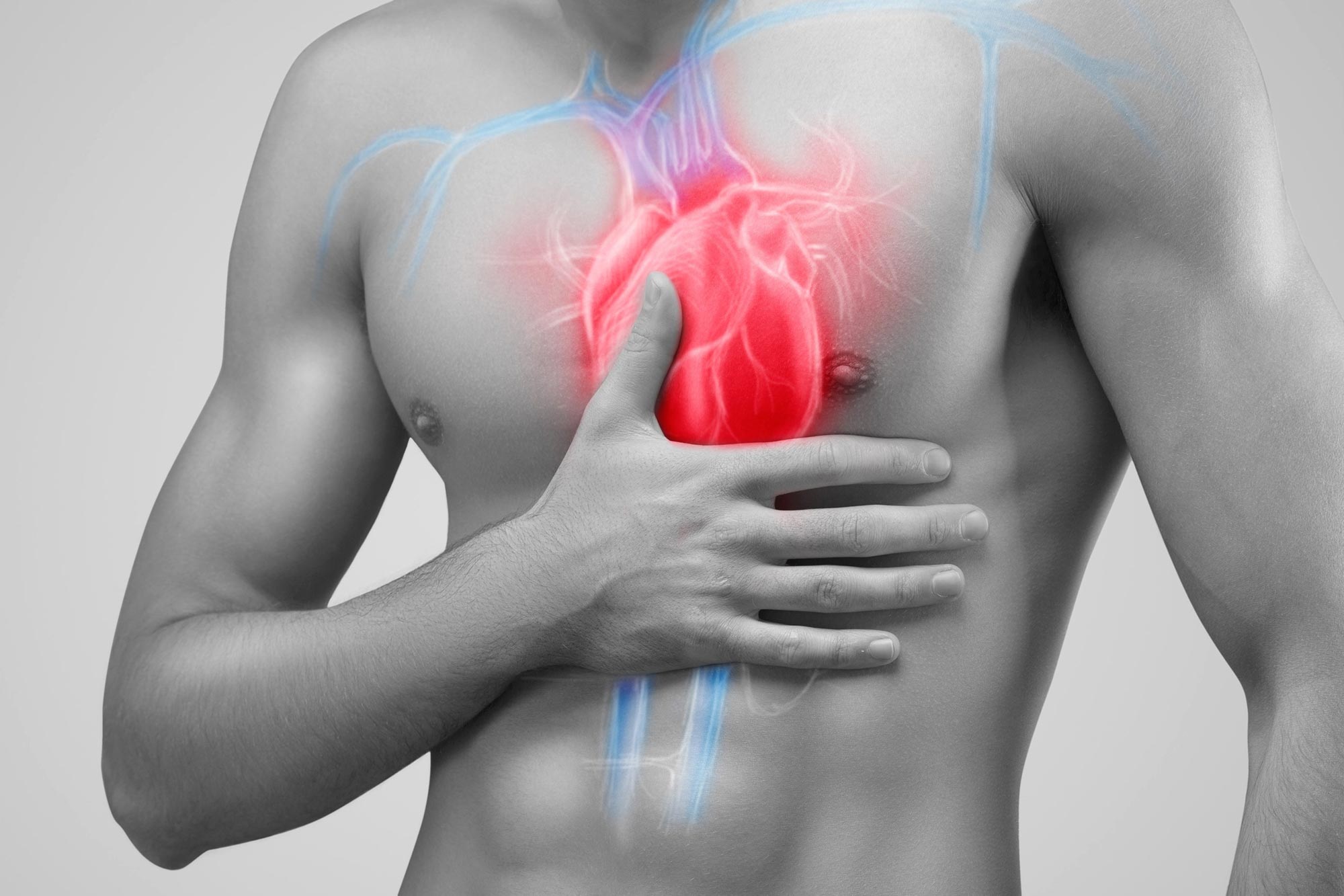
Revolutionary findings by UH researchers have the potential to become a powerful clinical method for treating heart disease.
A “powerful clinical strategy” for treating heart disease may result from the recent discovery.

Robert Schwartz, Hugh and Lillie Cranz Cullen Distinguished Professor of biology and biochemistry, led the studies published in the Journal of Cardiovascular Aging. Credit: University of Houston
University of Houston researchers have developed a groundbreaking technique that, in mice, not only restores the heart muscle cells after a myocardial infarction (or heart attack) but also helps the cells regenerate.
According to Robert Schwartz, Hugh Roy and Lillie Cranz Cullen Distinguished Professor of biology and biochemistry at the UH College of Natural Sciences and Mathematics, the ground-breaking discovery, which was published in the Journal of Cardiovascular Aginghas the potential to develop into a powerful clinical strategy for treating heart disease in people.
The innovative method used by the study team delivers mutant transcription factors, which are proteins that regulate the conversion of[{” attribute=””>DNA into RNA, to mice hearts using synthetic messenger ribonucleic acid (mRNA).
“No one has been able to do this to this extent and we think it could become a possible treatment for humans,” said Schwartz, who led the study with recent Ph.D. graduate Siyu Xiao and Dinakar Iyer, a research assistant professor of biology and biochemistry.
Synthetic mRNA contributes to stem cell-like growth
The researchers demonstrated that two mutated transcription factors, Stemin and YAP5SA, work in tandem to increase the replication of cardiomyocytes, or heart muscle cells, isolated from mouse hearts. These experiments were conducted in vitro on tissue culture dishes.
“What we are trying to do is dedifferentiate the cardiomyocyte into a more stem cell-like state so that they can regenerate and proliferate,” Xiao said.
Stemin turns on stem cell-like properties from cardiomyocytes. Stemin’s crucial role in their experiments was discovered by Iyer, who said the transcription factor was a “game changer.” Meanwhile, YAP5SA works by promoting organ growth that causes the myocytes to replicate even more.

The figure shows an example of a repair of an infarcted adult mouse heart in vivo, made possible through injections of a combination of STEMIN and YAP5SA mRNA. After four weeks, sectioned hearts stained red showed a significant reduction of infarcted area Credit: The Journal of Cardiovascular Aging
In a separate finding published in the same journal, the team will report that Stemin and YAP5SA repaired damaged mouse hearts in vivo. Notably, myocyte nuclei replicated at least 15-fold in 24 hours following heart injections that delivered those transcription factors.
Bradley McConnell, professor of pharmacology, and graduate student Emilio Lucero in the UH College of Pharmacy collaborated on the study by producing the infarcted adult mouse model.
“When both transcription factors were injected into infarcted adult mouse hearts, the results were stunning,” Schwartz said. “The lab found cardiac myocytes multiplied quickly within a day, while hearts over the next month were repaired to near normal cardiac pumping function with little scarring.”
An added benefit of using synthetic mRNA, according to Xiao, is that it disappears in a few days as opposed to viral delivery. Gene therapies delivered to cells by viral vectors raise several biosafety concerns because they cannot be easily stopped. mRNA-based delivery, on the other hand, turns over quickly and disappears.
A Limited Number of Cardiomyocytes
Schwartz and Iyer worked on this study for several years, and Xiao focused on this research throughout her doctoral studies at UH. She graduated in the fall of 2020.
“I feel honored and lucky to have worked on this,” Xiao said. “This is a huge study in heart regeneration especially given the smart strategy of using mRNA to deliver Stemin and YAP5SA.”
The findings are especially important because less than 1% of adult cardiac muscle cells can regenerate. “Most people die with most of the same cardiomyocytes they had in the first month of life,” she said. When there is a heart attack and heart muscle cells die, the contracting ability of the heart can be lost.
Reference: “Mutant SRF and YAP synthetic modified mRNAs drive cardiomyocyte nuclear replication” by Siyu Xiao, Rui Liang, Azeez B. Muili, Xuanye Cao, Stephen Navran, Robert J. Schwartz and Dinakar Iyer, 19 May 2022, The Journal of Cardiovascular Aging.
DOI: 10.20517/jca.2022.17
The study was funded in part through the University of Houston, a Cullen Endowed Chair, the Texas Higher Education Coordinating Board, Leducq Foundation, and a sponsored research agreement from Animatus Biosciences, LLC.